About clubfoot
Clubfoot, or talipes equinovarus, is a treatable birth defect that affects approximately 150,000-200,000 children each year. Clubfoot is the most common musculoskeletal birth deformity.
It is a congenital condition, meaning that when it occurs it is always present at birth. One or both feet may be affected, and about 50 percent are bilateral cases. Clubfoot is now generally thought to develop after the first trimester of pregnancy and can sometimes be picked up on ultrasound from about 20 weeks.
What is Clubfoot?
Clubfoot causes the foot to turn inward and point downward. Shortened tendons and ligaments on the inside of the lower leg restrict outward movement and cause the foot to turn inward. Tight Achilles tendons cause the foot to point downward. Most children born with clubfoot are not missing any bones, muscles, or connective tissue.
Parents will know at birth if their child has clubfoot because the foot will be twisted inward and rigid. Some cases are diagnosed before birth during a routine ultrasound. If you are wondering if your child has clubfoot, contact a specialist doctor who has experience in diagnosing this condition (not all paediatricians know how to diagnose clubfoot).
What is the incidence of clubfoot?
The worldwide average rate of occurrence is about one per 1000 births (some sources say 1:750). Studies report that for Southern and East African blacks the occurrence is higher (2 to 3 per 1000 births)*, affecting about 1 in 500 children born in the region. There are almost 11,000 children born with clubfoot in Southern Africa every year, around 2,000 of them in South Africa. It occurs about twice as frequently in boys.
The congenital clubfoot appears to be of genetic origin, but no one is entirely sure what causes it. Most cases are idiopathic (unknown cause). Some families report having a history of it, but in others it is apparently an isolated case. If either of the parents were born with clubfoot, there is a higher chance that their children will have it; if one parent is affected with clubfoot, there is a three to four percent chance that the offspring will also be affected. When both parents have clubfoot, their children have a 15% chance of developing clubfoot. In one study it was concluded that if a family already has one baby with clubfoot, the chance of having another increases to 1 in 35.
The condition is not painful for the newborn, but walking with an uncorrected clubfoot can be very painful and difficult, if not impossible.
Many older children live with the burden and stigma of neglected or relapsed clubfoot. Neglected clubfoot has a long-term impact on a child’s quality of life. Children in rural areas or depressed socio-economic conditions are more affected by the lack of access to adequate care or information. These children face a life of disability due to untreated clubfoot.
The Solution to Clubfoot
Clubfoot must be corrected in order for the child to walk properly. It is not difficult to correct. However, which treatment to use has been a subject of controversy for over 150 years. Although surgery became more popular than manipulation and serial casting during the 1950s, towards the end of the 20th Century, there was a growing demand for non-surgical techniques, particularly the Ponseti Method, as it became better publicised. The Ponseti method is now the international standard of best practice for clubfoot treatment, endorsed by all major paediatric orthopaedic associations worldwide.
Over sixty years ago Dr Ignaçio Ponseti developed a method for treating clubfoot that requires the use of full-length plaster casts and special protocol. The ‘Ponseti Method’ consists of using a series of casts, gentle manipulation and the use of a special brace. This treatment is over 95% effective and it is the most cost effective treatment with no side effects. Treatment should start soon after birth. In infants the method takes about 4-6 weeks of plaster casts, changed weekly. For older children or complex cases more casts are usually required.
After correction, clubfoot deformity has a tendency to recur. To prevent recurrence, a foot abduction brace must be worn full-time for 3 months and then at night until age four. The clubfoot brace consists of shoes attached to an adjustable bar at a specific width and angle. The brace helps to retain the feet in the corrected position and is the most effective way to avoid recurrence.
Published papers report that the Ponseti Method is effective for older children, even in cases of failed surgery. The Ponseti Method is very cost effective and various healthcare providers can perform the treatment, making the Ponseti method a practical option for eradicating clubfoot.
“A well-conducted orthopaedic treatment, based on a sound understanding of the functional anatomy of the foot and on the biological response of young connective tissue and bone to changes in direction of mechanical stimuli [correct manipulation of the foot ligaments, joint capsules, and tendons followed by casting in the new position] can gradually reduce or almost eliminate these deformities in most clubfeet. Less than five per cent of infants with very severe, short, fat feet with stiff ligaments unyielding to stretching will need surgical correction. The parents of all other infants may be reassured that their baby, when treated by expert hands, will have a functional, plantigrade foot which is normal in appearance, requires no special shoes, and allows fairly good mobility.”
*References:
- Incidence and Patterns of Congenital Talipes Equinovarus (Clubfoot) Deformity At Queen Elizabeth Central Hospital, Banter, Malawi. 1Mkandawire N.C. MCh (ORTH); FCS (ECSA), 2Kaunda E Cert. NMT.
- Common birth defects in South African blacks. Kromberg JG, Jenkins T S Afr Med J 1982; 62: 599-602
- Congenital musculoskeletal malformations in South African blacks: a study on incidence.
Pompe Van Meerdervoort HF. S Afr Med J 1996; 50: 1853-5.
About clubfoot
Clubfoot has been a recognised condition since ancient Egypt. Pharaohs Siptah and Tutankhamun had clubfoot, Hippocrates and the Aztecs described the condition.
Clubfoot is the most common musculoskeletal birth deformity affecting 200 000 babies each year, but many people are not even aware of the condition, what causes it, or how it is treated. Studies report that the Southern and East African population have almost twice the world average incidence of clubfoot. There are almost 11 000 children born with clubfoot in Southern Africa every year.
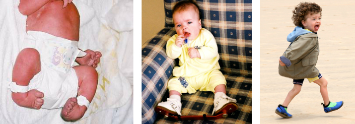
Clubfoot causes children to be born with one or both feet turned inward and upward. The foot is usually short and broad, curved in a bean shape with a deep crease on the sole because of the curve. The heel feels spongy because the tight Achilles tendon pulls the heel bone in. The calf muscles are often smaller and less developed. Without treatment the top of the foot is where the bottom should be, and the feet become fixed in this position, which makes walking either impossible or very painful.
Many older children live with the burden and stigma of neglected or relapsed clubfoot. Neglected clubfoot has a long-term impact on a child’s quality of life. Children in rural areas or depressed socio-economic conditions are more affected by the lack of access to adequate care or information. These children face a life of disability due to untreated clubfoot.
The Ponseti Method is over 95% effective when properly applied by a trained health care provider and is considered the “gold standard” treatment, leading to a normal, productive life.
Correcting clubfoot
The Ponseti method, devised over 60 years ago by Dr Ignacio Ponseti, avoids cutting the tight ligaments, tendons and joint capsules. It is a carefully constructed sequence of plaster casts and braces for children with clubfoot, based on his studies of the condition. Long-term follow-up studies show superior results to operative techniques.
Typically the method takes about four to six weeks of plaster casts, changed every five to seven days. For over 80% of cases, the tight Achilles tendon is cut in a minor procedure (tenotomy) and the corrected foot is put in a holding cast for three weeks to allow the tendon to regenerate longer.
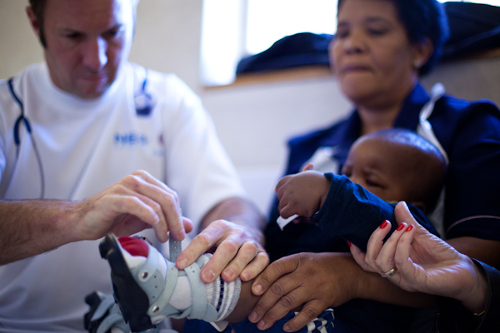
When the final cast is removed, a clubfoot brace is fitted – a pair of shoes attached to an adjustable bar at a specific width and angle. The child wears these at night for four years.
Clubfoot in Southern Africa
Clubfoot Treatment
If the Ponseti Method is done correctly, most clubfeet are corrected within four to six casting sessions. The casts are changed weekly. Less than 5% of clubfeet may be very stiff and severe; they may need more casting, but Dr Ponseti wrote that even they should be corrected within eight to 10 casting sessions. For the most resistant cases, surgery is sometimes required, but it’s less radical than it would have been without the correct casting method.
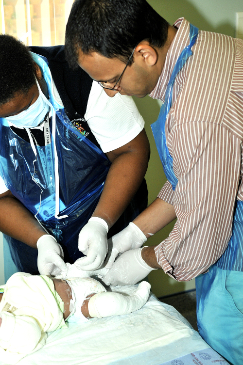
Dr Ponseti’s method uses gentle manipulations while the baby is seated comfortably on the mother’s lap. Each time, the manipulation is done slightly differently to stretch another part of the foot and ankle. Then the plaster is applied. While one person holds the foot in the required position, an assistant rolls on the plaster cast. A lot of plaster is wrapped around the knee, which is bent at almost a 90 degree angle. The Ponseti method requires a full-leg cast up to the groin. The position of the knee and the full leg cast helps to immobilise the foot into the correct position.
The cast is then left on for five to seven days to hold the correction achieved and allow the baby’s ligaments and tendons to soften into the new position. The cast is removed and the next manipulation is done and the foot and leg re-casted until the displaced bones are brought into the correct alignment and the foot is correctly positioned.
This is because the tissues forming the ligaments, joint capsules and tendons are still very elastic and stretch easily with each manipulation. It is also easier to cast a young baby than older child. However, Dr Ponseti and other doctors have successfully treated older babies and children, and avoided major surgery on the foot.
In many cases, before applying the last plaster cast, the Achilles tendon is cut in the doctor’s rooms. This is a simple procedure, which can be done with a local anaesthetic – it is a tiny cut at the back of the heel and no stitch required. By the time the cast is removed after three weeks, the tendon has regenerated to a proper length. The foot should appear overcorrected at first; this will change over time as the baby starts walking.
Following correction, the congenital condition that caused the clubfoot deformity in the first place tends to stay active and the foot can sometimes relapse. To prevent recurrence, when the last plaster cast is removed a foot abduction brace must be worn full-time, usually for three months and thereafter at night until four years of age. The clubfoot brace consists of a bar, with shoes attached to the ends of the bar. The gap between the shoes should be set to shoulder width, at 60-70º of external rotation and slightly angled up (toe higher than heel) at 10-15º of dorsiflexion to maintain the Achilles tendon length correction. In children with only one clubfoot, the shoe for the non-clubfoot is fixed on the bar at 30-40º of external rotation. During the daytime the children are barefoot or wear regular shoes. No stretching of the foot or physiotherapy is usually required, but some Ponseti practitioners give stretching exercises to be done at home.
The doctor can feel the position of the bones and the degree of correction, so X-rays of the feet are not required for young babies.
If the clubfoot brace wear is adhered to completely according to the recommended schedule, clinics report a 95% success rate. Some resistant foot may have recurrence, in which case further casting is done followed by a TATT or ATTT, a tibialis anterior tendon transfer. This is only done when the child is 3 to 4 years of age. The surgery consists of transferring the anterior tibial tendon to the third cuneiform, acting like an internal splint. This procedure does not have the negative after effects of full clubfoot surgery (Postero Remedial Release – PMR), such as scar tissue causing early onset arthritis.
Dr Ponseti’s opinion was that the poor results of cast and manipulative treatments of clubfeet by some doctors indicate that the attempts at correction have been inadequate because the techniques used are flawed.
Without a thorough understanding of the anatomy and kinematics of the normal foot and of the deviation of the bones in the clubfoot, the deformity is difficult to correct. Poorly conducted manipulations and casting further compound the clubfoot deformity rather than correct it, making treatment difficult or impossible.
Surgeons with limited experience in the treatment of clubfoot should not attempt to correct the deformity. They may succeed in correcting mild clubfeet, but the severe cases require experienced hands.
Referral to a doctor with training and expertise in the Ponseti non-surgical correction of clubfoot should be sought before considering surgery.